


Retina
By: Administrator
Date Uploaded: 05/02/2019
Tags: Retina
Attachments: image.png (6KB) image.png (42KB) image.png (42KB) image.png (46KB) image.png (37KB) image.png (38KB) image.png (2KB) image.png (39KB)
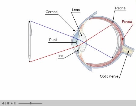
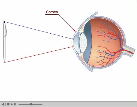
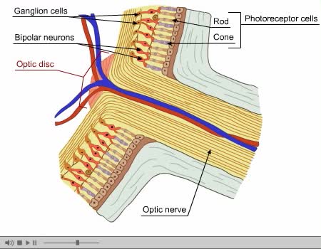
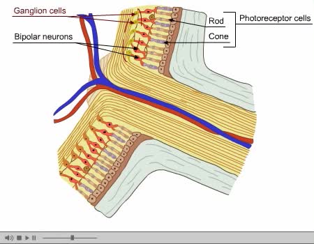
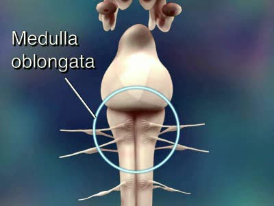
Eye Composed of special anatomical structures that work together to facilitate sight: Cornea Pupil Lens Vitreous body Light stimulates sensory receptors (rods and cones) in the retina or innermost layer of the eye. Vision is made possible through the coordinated actions of nerves that control: the movement of the eyeball the amount of light admitted by the pupil the focusing of that light on the retina by the lens the transmission of the resulting sensory impulses to the brain by the optic nerve. Eyeball's Outer Layer Composed of: Sclera, or white of the eye Cornea, or transparent anterior portion of the eye's fibrous outer surface. The curved surface of the cornea is important because it bends light rays and helps to focus them on the surface of the retina. Middle Layer: Choroid A pigmented vascular membrane that prevents internal reflection of light. Inner Layer The retina contains photoreceptive cells (rods and cones) that translate light waves into nerve impulses. Inner Layer The eye contains approximately 120 million rods that are sensitive to dim light. The rods contain rhodopsin, a pigment necessary for night vision. Optic disk: the point at which nerve fibers from the retina converge to form the optic nerve.
Add To
You must login to add videos to your playlists.
Advertisement
Suggestions

Comments
0 Comments total
Sign In to post comments.
No comments have been posted for this video yet.