Muscle cell structures - actin, myosin and titin filaments
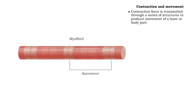
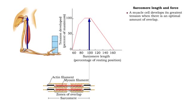
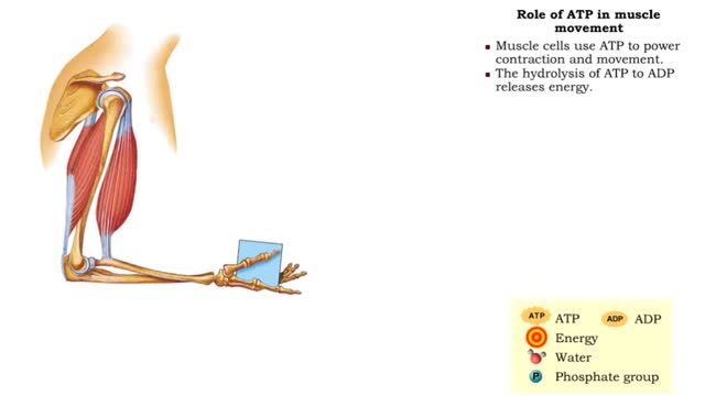
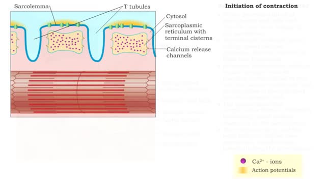
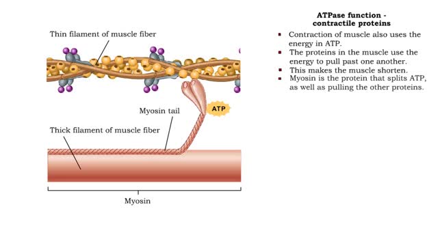
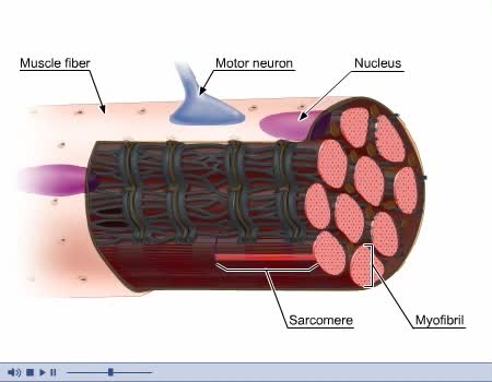
Once the muscle cell has been excited it will contract. • A muscle action potential will trigger the release Of Ca2+ ions into the sarcoplasm. • The Ca2+ ions bind to the regulatory proteins and trigger contraction. • Within skeletal muscle cells are structures that provide the ability to contract forcefully. • Myofibrils: rod-like structures that extend the length of the cell and contain repeating units called sarcomeres. • Sarcomeres: contractile units of a skeletal muscle cell that contain actin (thin) and myosin (thick) filaments. • When these filaments move over each other, the muscle cell contracts. ☆ Muscle cell structures - actin filament • The actin filament consists of actin proteins and regulatory proteins. • The actin protein has binding sites for myosin molecules. • The regulatory protein, tropomyosin, is wrapped around the actin molecules, covering the myosin binding sites. • A second regulatory protein, troponin, contains binding sites for Ca2+ ions and is attached to actin and tropomyosin. • In the presence of Ca2+ ions, troponin binds with calcium and pulls the tropomyosin away from the myosin binding sites and uncovers them. ☆ Muscle cell structures - myosin filament • The myosin filament consists of myosin molecules, each with a head (or crossbridge) and tail. • ATP binds to the crossbridge. • When ATP binds with myosin it is hydrolyzed, releasing energ to move the myosin protein. • Myosin crossbridges attach to specific myosin binding areas. • When the actin and myosin binds, any movement of the myosin filament will also move the actin filament. ☆Muscle cell structures - titin filament •The elastic titin filament allows the sarcomere to return to its resting length after contracting or stretching. Category
Add To
You must login to add videos to your playlists.
Advertisement


Comments
0 Comments total
Sign In to post comments.
No comments have been posted for this video yet.