

Brain Anatomy Animation (Part 2 of 2)
By: Administrator
Date Uploaded: 05/02/2019
Tags: Human brain Gross anatomy Blood supply Part 2
Attachments: image.png (20KB) image.png (26KB) image.png (27KB) image.png (46KB) image.png (36KB) image.png (46KB) image.png (52KB) image.png (33KB) image.png (40KB) image.png (55KB) image.png (55KB) image.png (54KB) image.png (23KB) image.png (17KB) image.png (46KB) image.png (22KB) image.png (48KB) image.png (18KB) image.png (18KB) image.png (42KB) image.png (39KB) image.png (59KB) image.png (38KB) image.png (46KB)
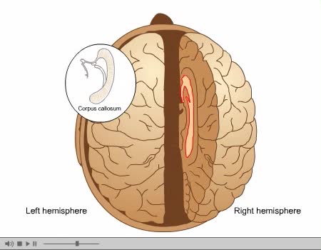
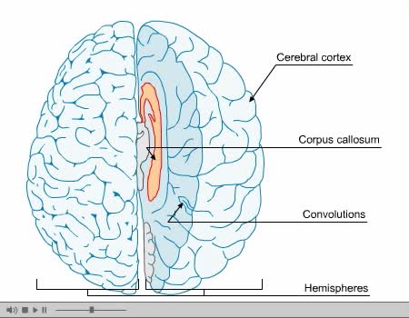
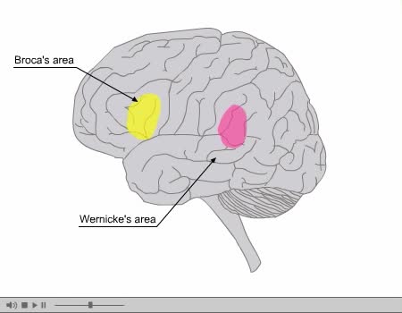
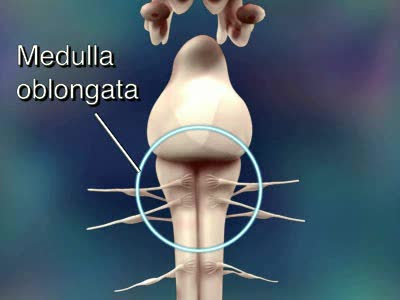
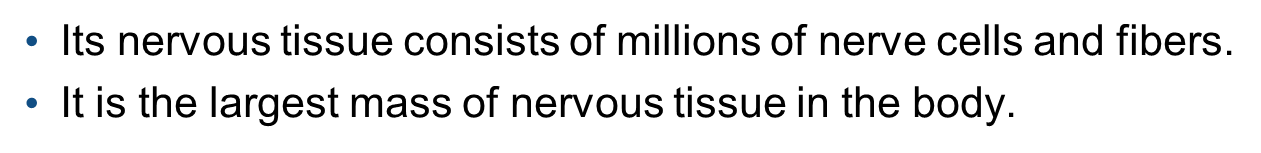
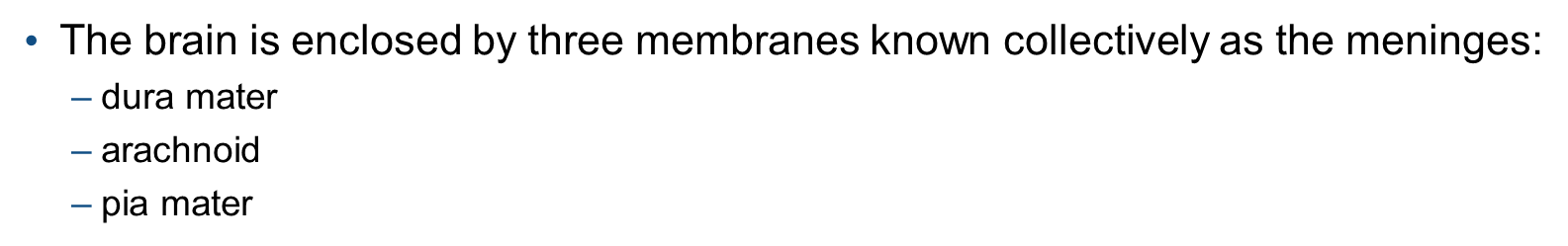
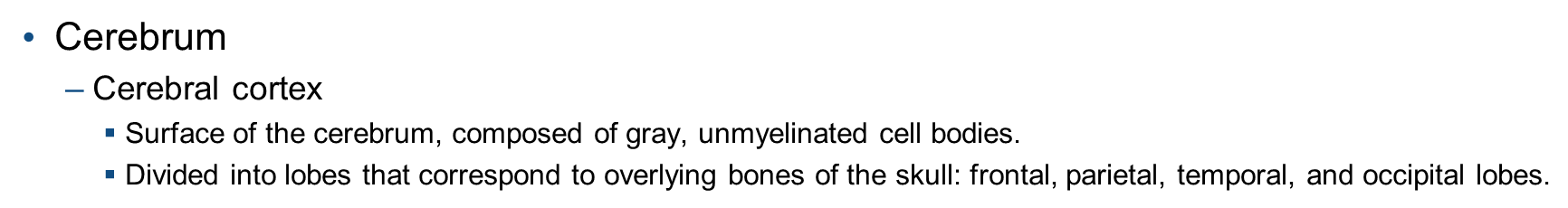
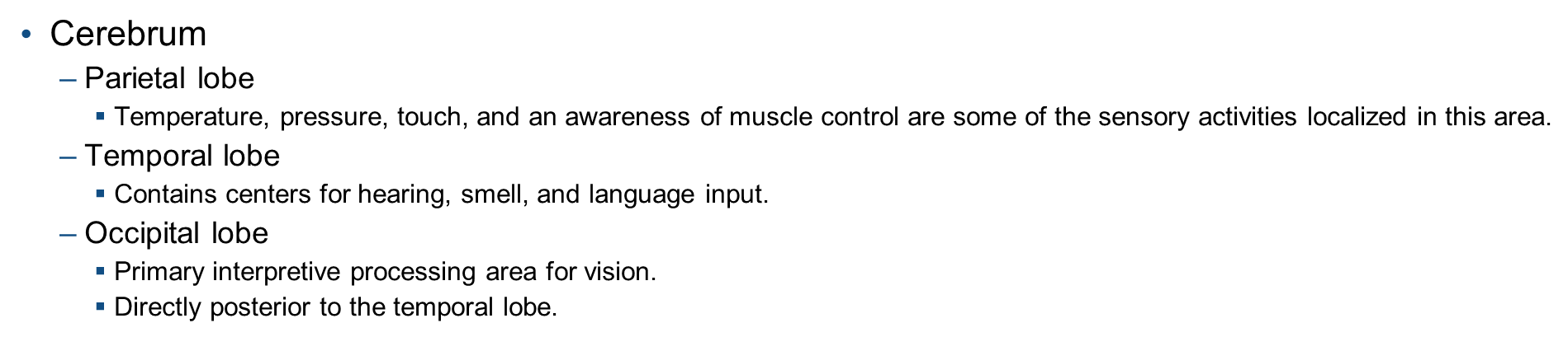
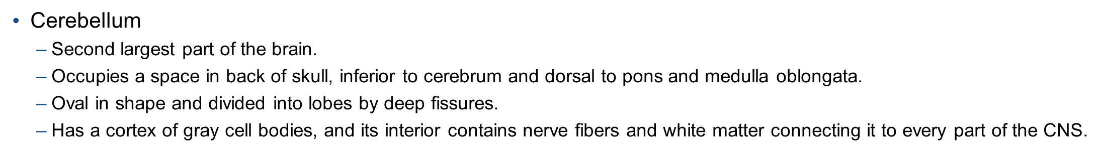
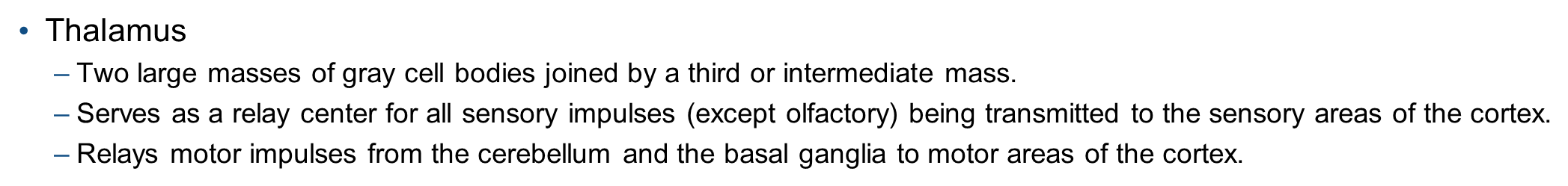
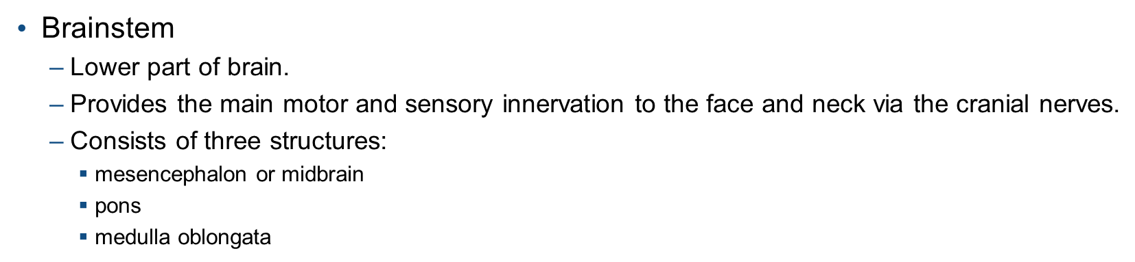
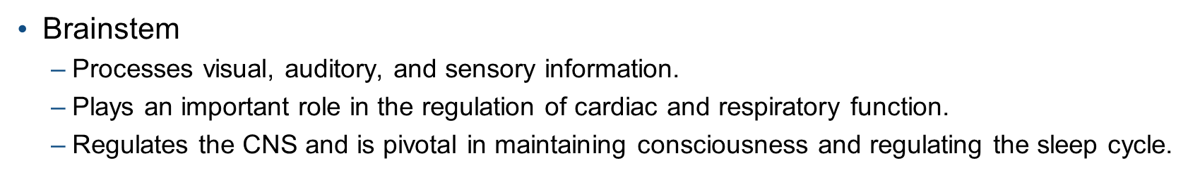
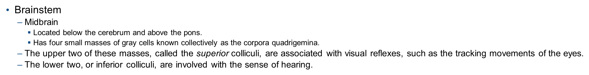
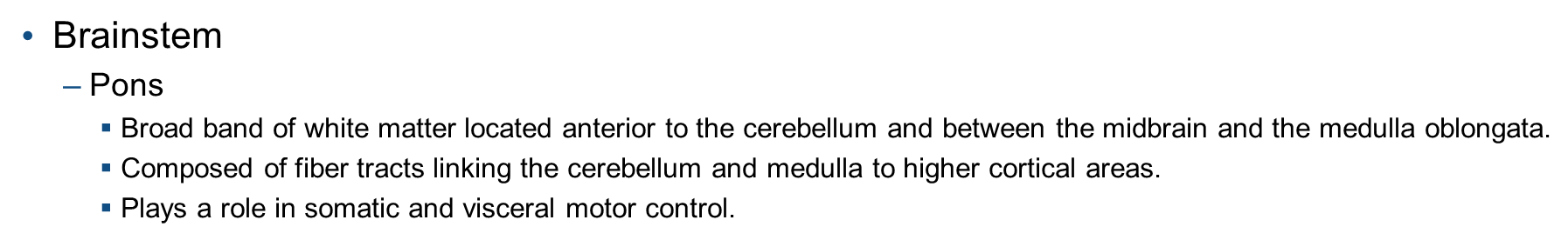
Its nervous tissue consists of millions of nerve cells and fibers. It is the largest mass of nervous tissue in the body. The brain is enclosed by three membranes known collectively as the meninges: dura mater arachnoid pia mater The major structures are the: cerebrum cerebellum diencephalon brainstem midbrain pons medulla oblongata A baby's brain has three main structural parts: cerebrum control center that receives, processes, and acts on information. cerebellum helps coordinate muscle activities and maintains posture and balance. brainstem maintains vital body functions, such as heartbeat, blood pressure, digestion, and swallowing. In an infant, the right and left hemispheres of the brain have yet to develop their own specific tasks. By the time a child is 3 years old, the two sides of the brain are well on their way to becoming specialized for different tasks. Cerebrum Represents 7/8 of brain's total weight. Contains nerve centers that evaluate and control all sensory and motor activity, including: sensory perception emotions consciousness memory voluntary movements Cerebrum Divided by the longitudinal fissure into two cerebral hemispheres, the right and left, that are joined by large fiber tracts (corpus callosum) that allow information to pass from one hemisphere to the other. Gyrus or convolution: Each bulge of the cerebrum. Sulcus: Each shallow furrow of the cerebrum. Cerebrum Cerebral cortex Surface of the cerebrum, composed of gray, unmyelinated cell bodies. Divided into lobes that correspond to overlying bones of the skull: frontal, parietal, temporal, and occipital lobes. Cerebrum Frontal lobe: Brain's major motor area. Site for personality and speech. Parietal lobe: Contains centers for sensory input from all parts of the body. Known as the somesthetic area. Site for interpretation of language. Cerebrum Parietal lobe Temperature, pressure, touch, and an awareness of muscle control are some of the sensory activities localized in this area. Temporal lobe Contains centers for hearing, smell, and language input. Occipital lobe Primary interpretive processing area for vision. Directly posterior to the temporal lobe. Cerebrum Parietal lobe Temperature, pressure, touch, and an awareness of muscle control are some of the sensory activities localized in this area. Temporal lobe Contains centers for hearing, smell, and language input. Occipital lobe Primary interpretive processing area for vision. Directly posterior to the temporal lobe. Cerebellum Second largest part of the brain. Occupies a space in back of skull, inferior to cerebrum and dorsal to pons and medulla oblongata. Oval in shape and divided into lobes by deep fissures. Has a cortex of gray cell bodies, and its interior contains nerve fibers and white matter connecting it to every part of the CNS. Cerebellum Plays an important part in coordination of voluntary and involuntary complex patterns of movement and adjusts muscles to maintain posture. Diencephalon Means second portion of the brain and refers to the thalamus and hypothalamus. Thalamus Two large masses of gray cell bodies joined by a third or intermediate mass. Serves as a relay center for all sensory impulses (except olfactory) being transmitted to the sensory areas of the cortex. Relays motor impulses from the cerebellum and the basal ganglia to motor areas of the cortex. Thalamus Some impulses related to emotional behavior are also passed from the hypothalamus, through the thalamus, to the cerebral cortex. Hypothalamus Lies beneath the thalamus. Is a principal regulator of autonomic nervous activity that is associated with behavior and emotional expression. It also produces neurosecretions for the control of water balance, sugar and fat metabolism, regulation of body temperature, and other metabolic activities. Hypothalamus Pituitary gland is attached to the hypothalamus by a narrow stalk, the infundibulum. Brainstem Lower part of brain. Provides the main motor and sensory innervation to the face and neck via the cranial nerves. Consists of three structures: mesencephalon or midbrain pons medulla oblongata Brainstem Processes visual, auditory, and sensory information. Plays an important role in the regulation of cardiac and respiratory function. Regulates the CNS and is pivotal in maintaining consciousness and regulating the sleep cycle. Brainstem Midbrain Located below the cerebrum and above the pons. Has four small masses of gray cells known collectively as the corpora quadrigemina. The upper two of these masses, called the superior colliculi, are associated with visual reflexes, such as the tracking movements of the eyes. The lower two, or inferior colliculi, are involved with the sense of hearing. Brainstem Pons Broad band of white matter located anterior to the cerebellum and between the midbrain and the medulla oblongata. Composed of fiber tracts linking the cerebellum and medulla to higher cortical areas. Plays a role in somatic and visceral motor control. Brainstem Medulla Oblongata Connects the pons and the rest of the brain to the spinal cord. All afferent and efferent tracts from the spinal cord either pass through or terminate in the medulla oblongata. Contains nerve centers for regulation and control of breathing, swallowing, coughing, sneezing, vomiting, the heartbeat, and blood pressure.
Add To
You must login to add videos to your playlists.
Advertisement
Suggestions

Comments
0 Comments total
Sign In to post comments.
No comments have been posted for this video yet.