Search Results
Results for: 'sensory nerves'
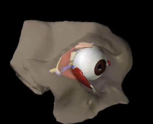
Structures of the Eye Animation
By: Administrator, Views: 2250
Orbit A cone-shaped cavity in the front of the skull that contains the eyeball. Formed by the combination of several bones and is lined with fatty tissue that cushions the eyeball. This cavity has several foramina (openings) through which blood vessels and nerves pass. Largest opening is the ...
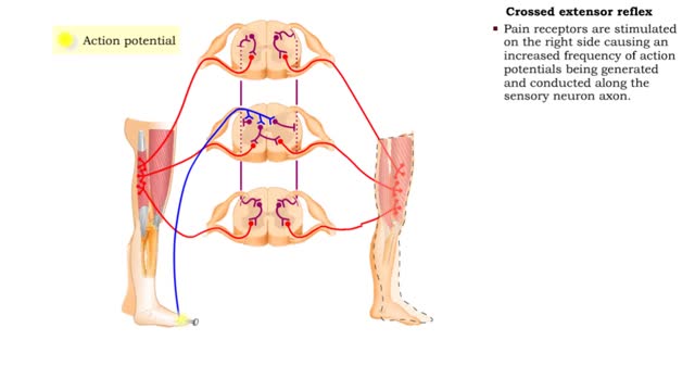
Flexor reflex & Crossed extensor reflex
By: HWC, Views: 10455
• The flexor reflex is a response to pain. This reflex is polysynaptic, ipsilateral, and intersegmental. • Pain receptors are stimulated causing increased frequency of action potentials to be generated and conducted along the sensory neuron axon. • The sensory impulses excite several ass...
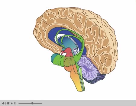
Brain Anatomy Animation (Part 2 of 2)
By: Administrator, Views: 14820
Its nervous tissue consists of millions of nerve cells and fibers. It is the largest mass of nervous tissue in the body. The brain is enclosed by three membranes known collectively as the meninges: dura mater arachnoid pia mater The major structures are the: cerebrum cerebellum dienc...
By: Administrator, Views: 13597
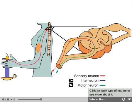
Interneurons: - Are called central or associative neurons. - Located entirely within the central nervous system. - They function to mediate impulses between sensory and motor neurons.
By: Administrator, Views: 13826
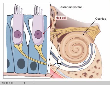
Process of Hearing Sound waves are directed to the eardrum, causing it to vibrate. These vibrations move the three small bones of the middle ear (malleus, incus, and stapes). Movement of stapes at oval window sets up pressure waves in the perilymph and endolymph. Process of Hearing The wav...
By: HWC, Views: 9827
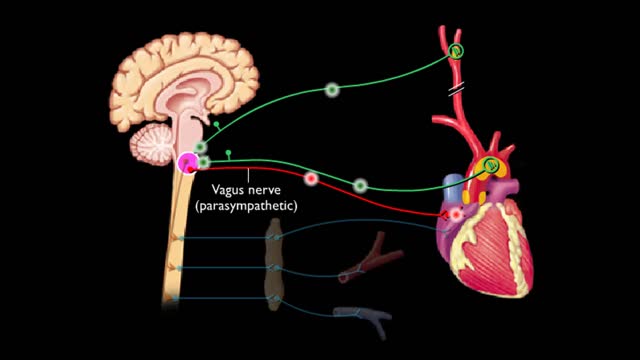
Baroreceptors located In the carotid sinus and the arch of the aorta respond to increases in blood pressure. Increased blood pressure stretches the carotid arteries and aorta causing the baroreceptors to increase their basal rate of action potential generation. Action potentials are conduct...
Membrane transport proteins - pores, gated channels and pumps
By: HWC, Views: 10689
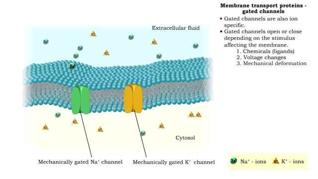
• a Three different types of membrane ion transport proteins are required to produce and carry electrical signals: • Pores • Gated channels • Na+/ K+ pump • Pores are always open and allow the diffusion of Na+ and K+ ions across the membrane, down their concentration gradients...
Optic Nerve and Optic Disk Animation (Part 2 of 2)
By: Administrator, Views: 13546
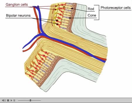
The optic disc or optic nerve head is the point of exit for ganglion cell axons leaving the eye. Because there are no rods or cones overlying the optic disc, it corresponds to a small blind spot in each eye. The ganglion cell axons form the optic nerve after they leave the eye. The optic disc ...
Advertisement


