


Brain Lobes Animation
By: Administrator
Date Uploaded: 05/02/2019
Tags: Brain anatomy lobes visual
Attachments: image.png (14KB)
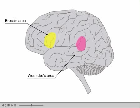
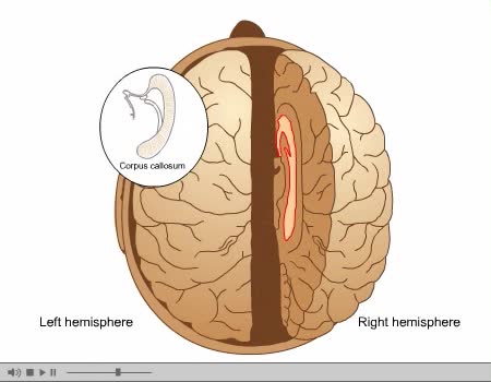
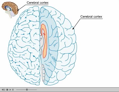
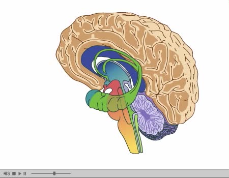
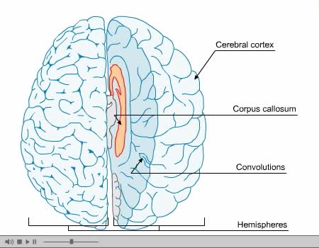
The brain’s cerebral cortex is the outermost layer that gives the brain its characteristic wrinkly appearance. The cerebral cortex is divided lengthways into two cerebral hemispheres connected by the corpus callosum. Traditionally, each of the hemispheres has been divided into four lobes: frontal, parietal, temporal and occipital. (Wikimedia) Although we now know that most brain functions rely on many different regions across the entire brain working in conjunction, it is still true that each lobe carries out the bulk of certain functions. Lobes of the brain QBI neuroscience (QBI) Bumps and grooves of the brain In humans, the lobes of the brain are divided by a number of bumps and grooves. These are known as gyri (bumps) and sulci (groves or fissures). The folding of the brain, and the resulting gyri and sulci, increases its surface area and enables more cerebral cortex matter to fit inside the skull. Image: (L) DJ / CC BY-SA 2.0 (R) Albert Kok / Public Domain Frontal lobe The frontal lobe is separated from the parietal lobe by a space called the central sulcus, and from the temporal lobe by the lateral sulcus. The frontal lobe is generally where higher executive functions including emotional regulation, planning, reasoning and problem solving occur. This is why in frontotemporal dementia, personality changes are often the first signs of the disease. The most famous case of frontal lobe dysfunction is the story of railway worker Phineas Gage. In 1848, Gage was using a tamping iron to pack in gunpowder for blasting a tunnel through rock. While his head was slightly turned, a mistaken strike sparked an explosion that forced the rod upwards into his left eye and out through his skull. Miraculously, Gage survived, blinded in his left eye and sustaining damage to much of his left frontal lobe. After the accident, others noticed changes in Gage’s personality: before the accident, he was known as responsible and hard-working, but afterwards, he became disrespectful, foul-mouthed and had difficulty carrying out plans. The frontal lobe also contains the primary motor cortex, the major region responsible for voluntary movement. Image: In 1848, Phineas Gage survived an explosion that drove a tamping iron through his left frontal lobe. (Image: Ratiu et al / CC BY-SA 2.1) Parietal lobe The parietal lobe is behind the frontal lobe, separated by the central sulcus. Areas in the parietal lobe are responsible for integrating sensory information, including touch, temperature, pressure and pain. Because of the processing that occurs in the parietal lobe, we are able to, for example, discern from touch alone that two objects touching the skin at nearby points are distinct, rather than one object. This process is called two-point discrimination. Different areas of the body have more sensory receptors, and so are more sensitive than others in discerning distinct points. Using callipers or a folded paperclip, and asking a subject to keep their eyes closed, this test can be used to check parietal lobe function. (Image: Lawrence House: Public Domain) While a subject's eyes are closed, a folded paperclip can be used to test two-point discrimination, which is mediated by the parietal lobe. The tester alternates using one point and two points on the area being tested (e.g. finger, shoulder, arm). The subject is asked to report whether they felt one or two points. Temporal lobe Separated from the frontal lobe by the lateral fissure, the temporal lobe also contains regions dedicated to processing sensory information, particularly important for hearing, recognising language, and forming memories. Auditory information The temporal lobe contains the primary auditory cortex, which receives auditory information from the ears and secondary areas, and processes the information so we understand what we’re hearing (e.g. words, laughing, a baby crying). Visual processing Certain areas in the temporal lobe make sense of complex visual information including faces and scenes. Memory The medial (closer to the middle of the brain) temporal lobe contains the hippocampus, a region of the brain important for memory, learning and emotions. Occipital lobe The occipital lobe is the major visual processing centre in the brain. The primary visual cortex, also known as V1, receives visual information from the eyes. This information is relayed to several secondary visual processing areas, which interpret depth, distance, location and the identity of seen objects.
Add To
You must login to add videos to your playlists.
Advertisement
Suggestions
Comments
0 Comments total
Sign In to post comments.
No comments have been posted for this video yet.