


A Human Karyotype Preparation Animation

By: HWC
Date Uploaded: 12/09/2021
Tags: A Human Karyotype Preparation Animation Colchicine mitosis centrifuge tube a hypotonic salt solution lysed cells the cell's chromosomes homologous chromosomes karyotype diagram metaphase chromosomes karyotype of a normal male
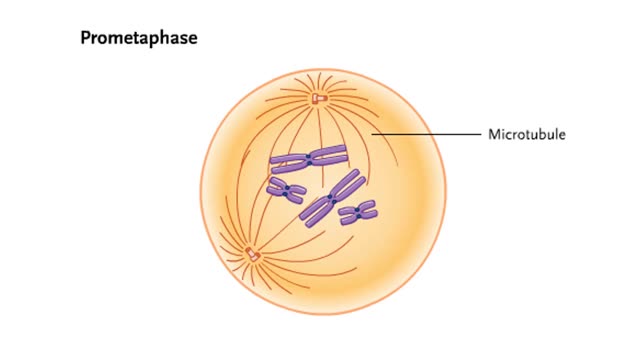
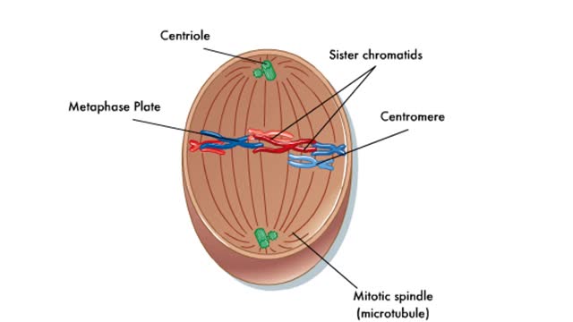
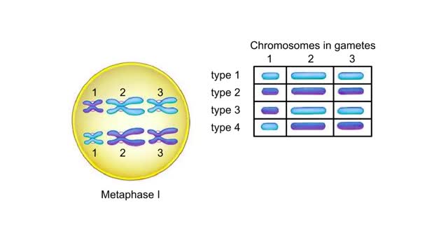
Blood is collected from the person being analyzed. The blood is added to a growth medium that also contains a chemical that stimulates mitosis. The cells are allowed to grow in this medium for two or three days at body temperature. Colchicine is added to arrest cell division at metaphase. The arrested cells are transferred to a centrifuge tube. Centrifugation concentrates the cells at the bottom of the tube. Addition of a hypotonic salt solution causes them to swell up and move apart. The lysed cells are prepared, fixed, and placed on a microscope slide. A cell is observed and photographed under the microscope. The part of the photo that shows the cell's chromosomes is enlarged and images of individual chromosomes are cut out. Finally, the images are arranged so that all pairs of homologous chromosomes are horizontally aligned by centromeres. The result is a karyotype diagram of the arrested metaphase chromosomes. This is the karyotype of a normal male.
Add To
You must login to add videos to your playlists.
Advertisement
Suggestions
Comments
0 Comments total
Sign In to post comments.
No comments have been posted for this video yet.