Search Results
Results for: 'Mechanisms for chromosome movement Animation'
By: HWC, Views: 9107
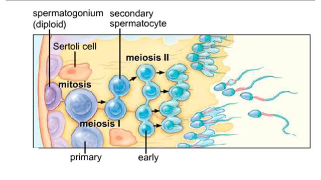
Spermatogenesis takes place inside the seminiferous tubules. Diploid spermatogonia located near the outer edge of the tubule divide mitotically to form primary spermatocytes. The first meiotic division produces secondary spermatocytes with a haploid number of duplicated chromosomes. T...
By: Administrator, Views: 423
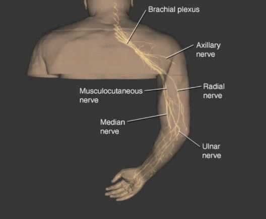
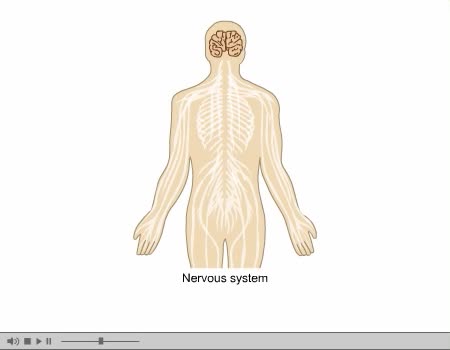
Used to describe neuronal processes conducting impulses from one location to another. Nerve fibers: - Nerve fibers of the PNS are wrapped by protective membranes called sheaths. - Myelinated fibers have an inner sheath of myelin, a thick fatty substance, and an outer sheath or neurilemma compo...
What are Taste Receptors? How Does it Work? Animation
By: HWC, Views: 7891
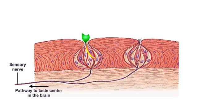
Do you ever wonder how you can taste the foods you eat? It all starts with taste receptors in your muscular tongue. Taste receptor neurons are found in your taste buds but you are not looking at the taste buds. The raised bumps on the surface of the tongue that you see are specialized epith...
Optic Nerve and Optic Disk Animation (Part 1 of 2)
By: Administrator, Views: 14084
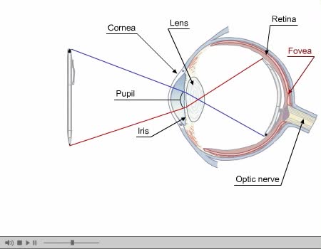
Inner Layer Blind spot: the absence of rods and cones in the area of the optic disk creates a blind spot on the retina's surface; the only part of the retina that is insensitive to light. Inner Layer The eye contains approximately 120 million rods that are sensitive to dim light. The rods ...
By: Administrator, Views: 395
Used to describe neuronal processes conducting impulses from one location to another. Nerve fibers: - Nerve fibers of the PNS are wrapped by protective membranes called sheaths. - Myelinated fibers have an inner sheath of myelin, a thick fatty substance, and an outer sheath or neurilemma compo...
By: Administrator, Views: 14012
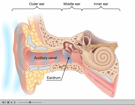
The ear is the organ of hearing and, in mammals, balance. In mammals, the ear is usually described as having three parts—the outer ear, the middle ear and the inner ear. The outer ear consists of the pinna and the ear canal. Since the outer ear is the only visible portion of the ear in most ani...
Vascular tissues in a corn stem and a buttercup root
By: HWC, Views: 5467
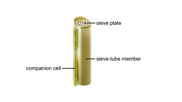
Vascular tissues in a corn stem and a buttercup root. The cells that make up each tissue. Xylem conducts water and dissolved ions. It also helps mechanically support a plant. The cells, called vessel members and tracheids, are dead at maturity. Their lignified walls interconnect and serve as p...
By: Administrator, Views: 13967
In the nervous system, a synapse is a structure that permits a neuron (or nerve cell) to pass an electrical or chemical signal to another neuron or to the target effector cell. Synapses are essential to neuronal function: neurons are cells that are specialized to pass signals to individual tar...
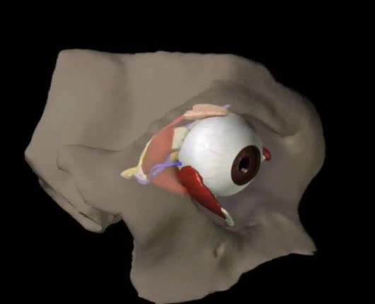
Structures of the Eye Animation
By: Administrator, Views: 2342
Orbit A cone-shaped cavity in the front of the skull that contains the eyeball. Formed by the combination of several bones and is lined with fatty tissue that cushions the eyeball. This cavity has several foramina (openings) through which blood vessels and nerves pass. Largest opening is the ...
Advertisement


