

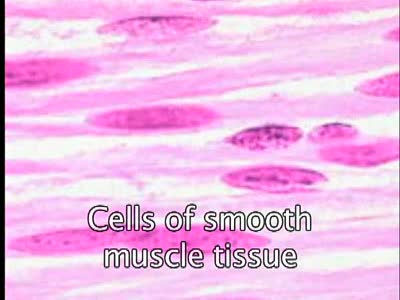
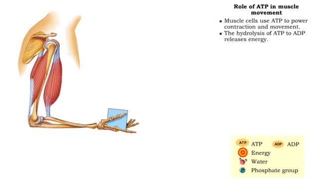
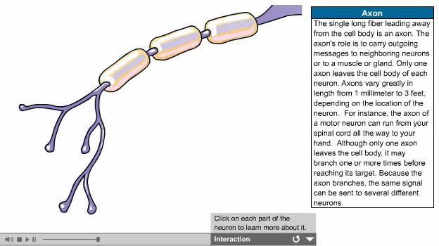
Nervous pathway to the Neuromuscular (NMJ)

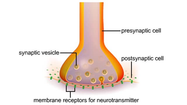
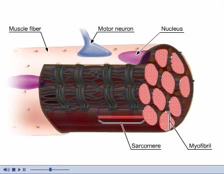
• A nervous impulse, also called an action potential, starts from the brain or spinal cord to signal skeletal muscle cell contraction. Action potentials continue along a motor neuron to the muscle cell. • The signal to contract must cross a synapse - the neuromuscular junction (NMJ) - between the neuron and the skeletal muscle cell. Components of the NMJ • The NMJ is a chemical synapse between the motor neuron membrane (synaptic end bulb) and the muscle cell membrane (sarcolemma). • The two cell membranes are separated by a physical space called the synaptic cleft. • The electrical nerve impulses (action potentials) cannot cross the synaptic cleft. • A neurotransmitter chemical (acetylcholine, in this case) must carry the information from cell to cell across the synapse. Neurotransmission at the NMJ • Action potentials arrive at the synaptic end bulb and alter the electrical voltage of the membrane. • This change in voltage opens voltage-gated calcium channels, allowing calcium to flood into the neuron. • Increased intracellular calcium causes synaptic vesicles (carrying acetylcholine) to fuse with the neuron membrane and acetylcholine is then released into the synaptic cleft. • The neurotransmitter acetylcholine binds to receptors (chemically gated sodium channels) on the sarcolemma. • The receptors open, and the large influx of sodium ions generates a muscle action potential along the sarcolemma. Initiation of contraction • The muscle action potential moves along the sarcolemma and continues down the T tubules and causes changes in the terminal cisterns of the sarcoplasmic reticulum (SR). • In a relaxed muscle, cisterns of the SR Store Ca2+ ions. • In response to the action potential, a chain of enzymatic reactions open ligand-gated calcium channels and release calcium ions into the cytosol. • Inside the cell, contractile proteins are arranged into contractile units called sarcomeres. • The sarcomeres are constructed of smaller structures called filaments. Relaxation • Once acetylcholine has propagated an action potential along the muscle cell membrane, it is destroyed by the enzyme acetylcholinesterase. • This prevents another muscle action potential from being generated except when a new neuron action potential causes the release of more acetylcholine. As action potentials no longer activate the Ca2 + ions release from the SR, Ca2+ ions are pumped back into the SR and stored until another action potential occurs.
Add To
You must login to add videos to your playlists.
Advertisement
Suggestions

Comments
0 Comments total
Sign In to post comments.
No comments have been posted for this video yet.