

Coaductile pathway, Timing of contraction signal & Conduction system and ECG

By: HWC
Date Uploaded: 11/29/2019
Tags: homeworkclinic.com Homework Clinic HWC Coaductile pathway SA node pacemaker Apex of the heart Purkinje fibers myofibers aorta pulmonary trunk Timing of contraction signal ventricle Conduction system and ECG electrocardiogram QRS waves atrial excitation
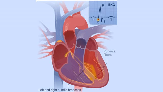
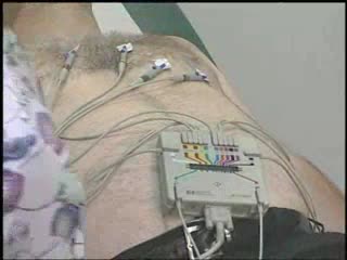
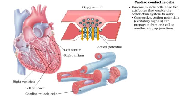
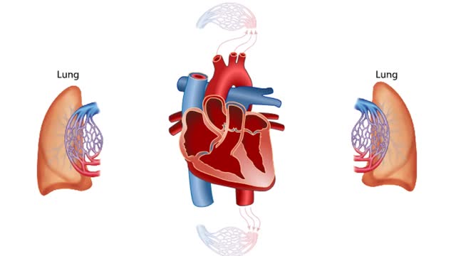
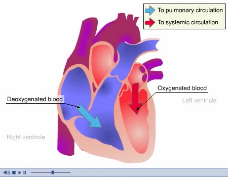
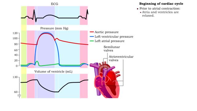
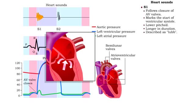
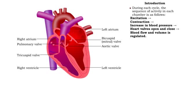
• When the system is healthy, the signal to contract the entire conduction system originates in the SA node - known as the heart's pacemaker. • The SA node triggers contraction because it depolarizes at a faster rate than other parts of the conduction system. • The wave of excitation from the SA node travels through the atria and triggers the atria to contract and move additional blood into the ventricles. ▪ The AV node is the only connection between the atria and ventricles. Once the signal passes the AV node, it propagates through the ventricular portion of the conduction system: • AV bundle and left and right bundle branches. • Apex of the heart. • Large-diameter conduction myofibers (Purkinje fibers). • The conduction myofibers then directly stimulate the cardiac cells in the ventricles to contract. • During ventricular contraction, blood is squeezed out from the apex to the base and into the pulmonary trunk and aorta. • For a resting heart, the signal travels its route from the SA node to the ventricles in less than a quarter-second. • The signal passes through the atria very quickly and then slows when it reaches the AV node. The slowing occurs because the AV node fibers have smaller diameters. • The delay allows the atria to fully contract, increasing blood volume in the ventricles before ventricle contraction begins. • The signal then quickly passes through the AV bundle and excites the apex and base of the ventricles, beginning contraction. • The conduction system is a series of electrical processes. The sequence of depolarizations and repolarizations can be seen in a normal electrocardiogram. (ECG). • The ECG can also reveal problems with the conduction system - a blockage, for example. • In this abnormal ECG trace, you can see that ventricular excitation (QRS waves) is independent of atrial excitation (P waves). • The pace of ventricular contraction is being determined by the ventricles' natural rhythm, not from excitation generated by the SA node. The SA nodes signals are not successfully moving along their normal path to the ventricles.
Add To
You must login to add videos to your playlists.
Advertisement
Suggestions

Comments
0 Comments total
Sign In to post comments.
No comments have been posted for this video yet.