

Electrical Conduction System of the Heart

By: HWC
Date Uploaded: 09/04/2020
Tags: homeworkclinic.com Homework Clinic HWC Electrical Conduction System of the Heart electrocardiogram EKG or ECG sinoatrial node SA node heart's right atrium P wave heart's atria atrioventricular (AV) node Q wave Purkinje fibers heart's ventricles R wave S wave pulmonary valve aortic valve T wave
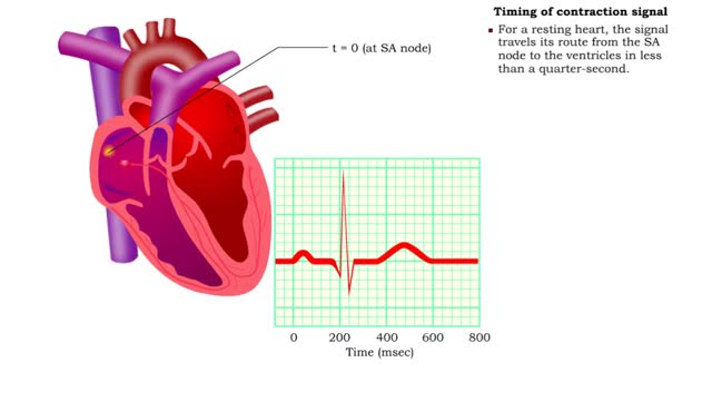
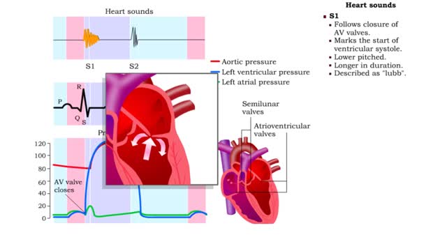
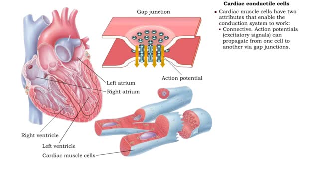
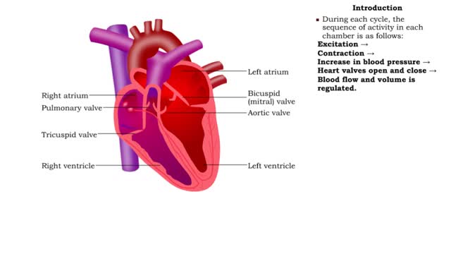
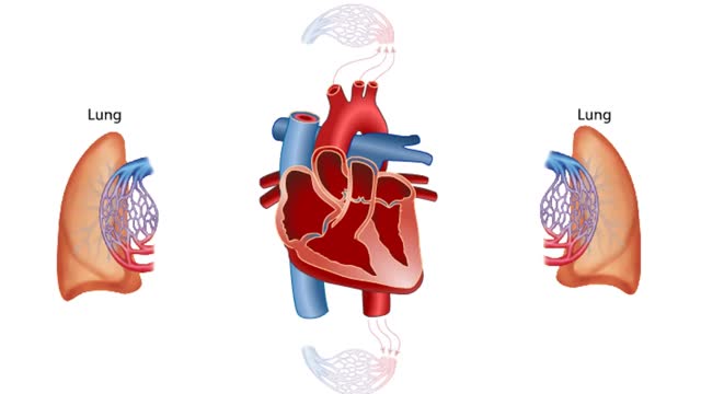
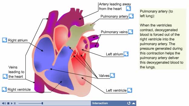
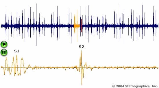
Your heart is a muscle that works continuously, much like a pump. Each beat of your heart is set in motion by an electrical signal from within your heart muscle. The electrical activity is recorded by an electrocardiogram. known as an EKG or ECG. Each beat of your heart begins with an electrical signal from the sinoatrial node. also known as the SA node. The SA node is located in your heart's right atrium. When your heart's right atrium is full with blood, the electrical signal spreads across the cells of your heart's right and left atria. This signal causes the atria to contract, or squeeze. This pumps blood through the open valves from the atria into both ventricles. The P wave on the EKG. marks the contraction of your heart's atria. The signal arrives at the atrioventricular (AV) node near the ventricles. Here it is slowed for an instant to allow your heart's right and left ventricles to fill with blood. On an EKG. this interval is represented by the start of the line segment between the P and Q wave. The signal is released and moves next to the bundle of His located in your heart's ventricles. From the bundle of His. the signal fibers divide into left and right bundle branches, which run through your heart's septum. On the EKG. this is represented by the Q wave. The signal leaves the left and right bundle branches through the Purkinje fibers that connect directly to the cells in the walls of your heart's ventricles. The signal spreads quickly across your heart's ventricles. As the signal spreads across the cells of the ventricle walls, both ventricles contract. but not at exactly the same moment. The left ventricle of your heart contracts an instant before the right ventricle. On an EKG, the R wave marks the contraction of your heart's left ventricle. The S wave marks the contraction of your heart's right ventricle. The contraction of your heart's right ventricle pushes blood through the pulmonary valve to your lungs. The contraction of your heart's left ventricle pushes blood through the aortic valve to the rest of your body. As the signal passes, the walls of your heart's ventricles relax and await the next signal. On the EKG, the T wave marks the point at which your heart's ventricles are relaxing. This process continue over and over.
Add To
You must login to add videos to your playlists.
Advertisement
Suggestions

Comments
0 Comments total
Sign In to post comments.
No comments have been posted for this video yet.