Cleavage and Implantation Animation
By: HWC
Date Uploaded: 11/01/2021
Tags: Cleavage and Implantation Animation Fertilization oviduct zygote uters mitotic divisions morula blastula blastocoel endometrium embryonic disk embryo amniotic cavity yolk sac blastocyst chorion placenta التخصيب
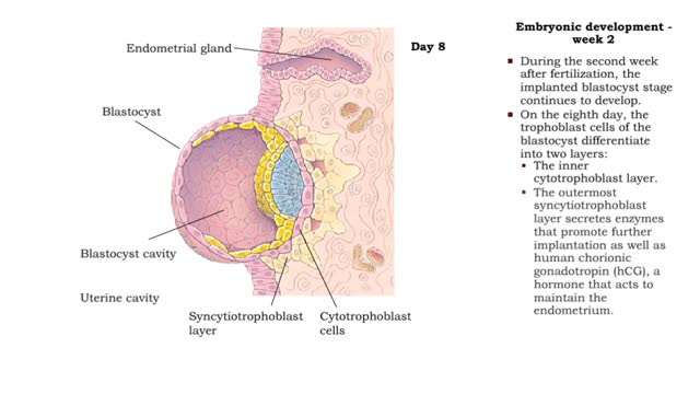
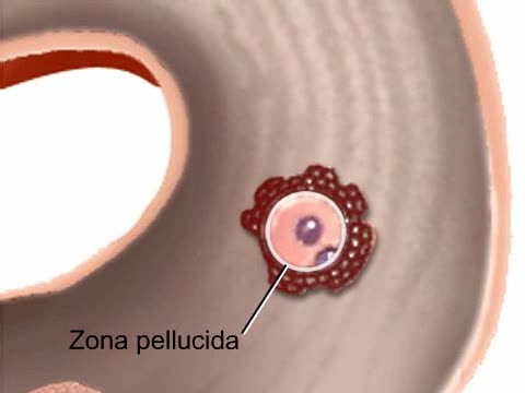
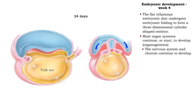
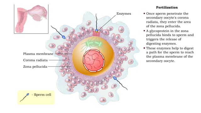
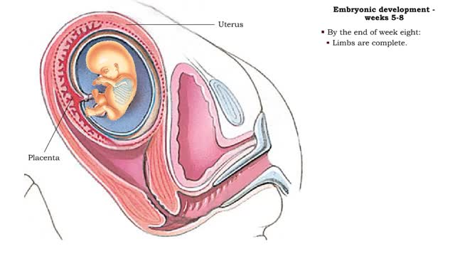
✔ https://HomeworkClinic.com ✔ https://Videos.HomeworkClinic.com ✔ Ask questions here: https://HomeworkClinic.com/Ask Follow us: ▶ Facebook: https://www.facebook.com/HomeworkClinic ▶ Review Us: https://trustpilot.com/review/homeworkclinic.com Fertilization typically takes place in the upper part of the oviduct. Cleavage begins as the zygote moves through the oviduct toward the uterus. Continued mitotic divisions produce a ball of sixteen to thirty-two cells called a morula. By the fifth day, a blastula has formed, with a surface layer of cells surrounding a fluid-filled blastocoel and an inner cell mass. About a week after fertilization, implantation is under way. The blastocyst adheres to the endometrium that lines the uterus and begins to send out projections into the maternal tissues. As implantation proceeds, the inner cell mass develops into an embryonic disk that is two cell layers thick. This will give rise to the embryo. Membranes start to form around the embryonic disk. The amniotic cavity will fill with fluid and cradle the embryo. The yolk sac will function in blood cell formation. Spaces in the maternal tissue around the implanting blastocyst open and fill with blood. Inside the blastocyst, a chorionic cavity opens around the amnion and yolk sac. The membrane that lines this cavity is the chorion. It will become part of the placenta.
Add To
You must login to add videos to your playlists.
Advertisement


Comments
0 Comments total
Sign In to post comments.
No comments have been posted for this video yet.