The Vestibular System
By: HWC
Date Uploaded: 07/28/2020
Tags: homeworkclinic.com Homework Clinic HWC the vestibular system temporal bone peripheral component labyrinth cochlea otolith organs utricle saccule vestibular hair cells cochlear hair cells ampullae sensory epithelium macula gelatinous layer otoconia kinocilium ampulla gelatinous mass endolymphatic fluid endolymph cupula
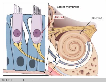
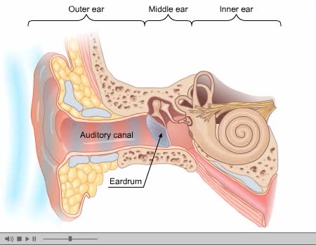
The vestibular system has important sensory and motor functions, contributing to the perception of self-motion, head position, and spatial orientation relative to gravity. The function of the vestibular system can be simplified by remembering some basic terminology of classical mechanics. All bodies moving in three dimensions have six degrees of freedom; three of these are translational and three are rotational. The translational components may be given in terms of movements along x, y, and z axes of the head. Rotations about the x, y, and z axes are commonly referred to as roll, pitch, and yaw. The positive direction of head rotation follows a right-hand rule—that is, if the fingers of the right hand are curled in the direction of the arrows, the thumb points in the positive direction of the axis. Buried deep in the temporal bone, the main peripheral component of the vestibular system is an elaborate set of interconnected chambers—the labyrinth—that has much in common, and is in fact continuous with, the cochlea. The labyrinth consists of the two otolith organs—the utricle and saccule—and three semicircular canals. The vestibular hair cells, which like cochlear hair cells transduce minute displacements into behaviorally relevant receptor potentials, are located in the utricle and saccule and in three juglike swellings called ampullae, located at the base of the semicircular canals next to the utricle. In the utricle and saccule, the sensory epithelium, or macula, consists of hair cells and associated supporting cells. Overlying the hair cells and their stereocilia is a gelatinous layer; above this layer is a fibrous structure, the otolithic membrane, in which are embedded crystals of calcium carbonate called otoconia. The crystals give the otolith organs their name (otolith is Greek for "ear stones"). The otoconia make the otolithic membrane considerably heavier than the structures and fluids surrounding it; thus, when the head tilts, gravity causes the membrane to shift relative to the sensory epithelium. The resulting shearing motion between the otolithic membrane and the macula displaces the hair bundles, which are embedded in the lower, gelatinous surface of the membrane. This displacement of the hair bundles generates a receptor potential in the hair cells that is dependent upon the direction of tilt. Movement of the stereocilia toward the kinocilium causes potassium channels to open, depolarizing the hair cell. The depolarization results in neurotransmitter release and excitation of the vestibular nerve fibers. Movement of the stereocilia in the direction away from the kinocilium closes the channels, hyperpolarizing the hair cell and thus reducing vestibular nerve activity. A shearing motion between the macula and the otolithic membrane also occurs when the head undergoes linear accelerations. Hair bundle displacement occurs transiently in response to linear accelerations and tonically in response to tilting of the head. The orientation of the stereocilia bundles relative to the kinocilia is such that, given the utricle and saccule on each side of the body, there is a continuous representation of all directions of body movement Ultimately, variations in hair cell polarity within the otolith organs produce patterns of vestibular nerve fiber activity that, at a population level, can unambiguously encode head position and the forces that influence it. Whereas the otolith organs are primarily concerned with head translations and orientation with respect to gravity, the semicircular canals sense head rotations, arising either from self-induced movements or from angular accelerations of the head imparted by external forces. Each of the three semicircular canals has at its base a bulbous expansion called the ampulla, which houses the sensory epithelium, or crista, that contains the hair cells. The structure of the canals suggests how they detect the angular accelerations that arise through rotation of the head. The hair bundles extend out of the crista into a gelatinous mass, the cupula, that bridges the width of the ampulla, forming a viscous barrier through which endolymph cannot circulate. As a result, the relatively compliant cupula is distorted by movements of the endolymphatic fluid. When the head turns in the plane of one of the semicircular canals, the inertia of the endolymph produces a force across the cupula, distending it away from the direction of head movement and causing a displacement of the hair bundles within the crista. In contrast, linear accelerations of the head produce equal forces on the two sides of the cupula, so the hair bundles are not displaced. Each semicircular canal works in concert with the partner located on the other side of the head that has its hair cells aligned oppositely. There are three such pairs: one pair of horizontal canals, and the superior canal on each side working with the posterior canal on the other side—both are in the same plane. The orientation of the horizontal canals makes them selectively sensitive to rotation in the horizontal plane. More specifically, the hair cells in the canal towards which the head is turning are depolarized, while those on the other side are hyperpolarized. For example, when the head turns to the left, the cupula is pushed toward the kinocilium in the left horizontal canal, and the firing rate of the relevant axons in the left vestibular nerve increases. In contrast, the cupula in the right horizontal canal is pushed away from the kinocilium, with a concomitant decrease in the firing rate of the related neurons. If the head turns to the right, the result is just the opposite. This push-pull arrangement operates for all three pairs of canals; the pair whose activity is modulated is in the plane of the rotation, and the member of the pair whose activity is increased is on the side toward which the head is turning. The net result is a system that provides information about the rotation of the head in any direction.
Add To
You must login to add videos to your playlists.
Advertisement




Comments
0 Comments total
Sign In to post comments.
No comments have been posted for this video yet.