


Chromosome structural organization/ Mechanisms for chromosome movement Animation

By: HWC
Date Uploaded: 06/20/2022
Tags: homeworkclinic.com HWC Chromosome structural organization Mechanisms for chromosome movement Animation Sister chromatids of a metaphase chromosome how the chromosome is organized metaphase sister chromatids centromere super coiled array chromosomal proteins and DNA cylindrical fiber histones mitotic metaphase microtubules kinetochore microtubules anaphase beam of ultraviolet light nonkinetochore microtubules
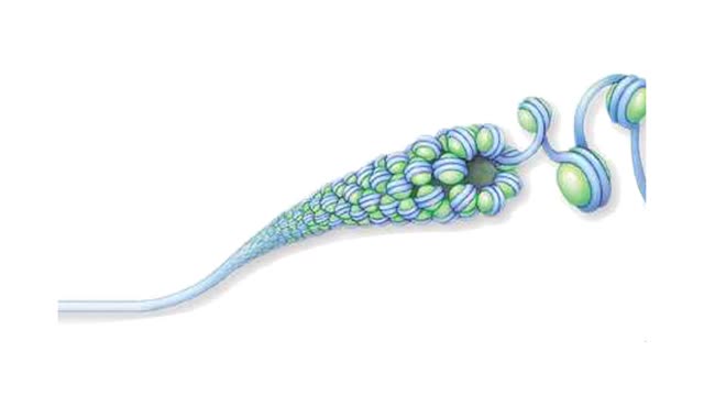
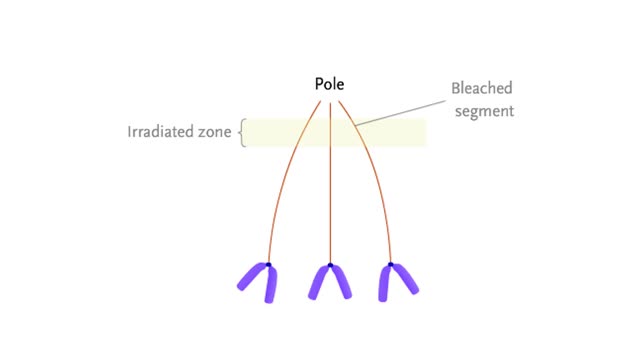
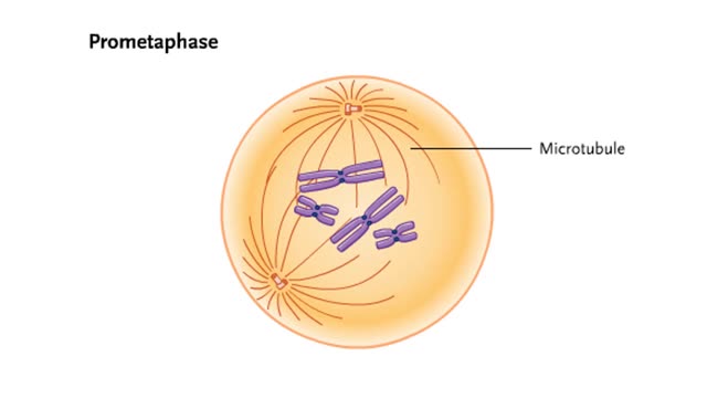
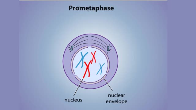
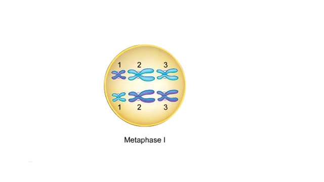
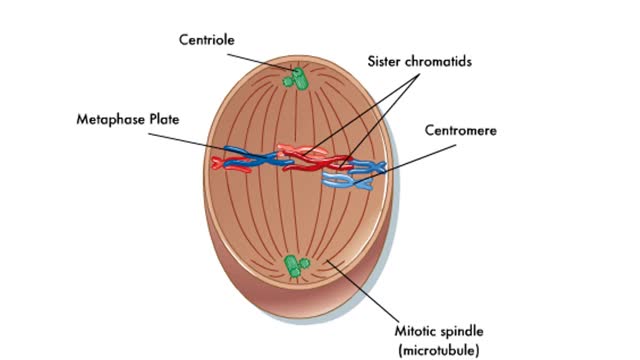
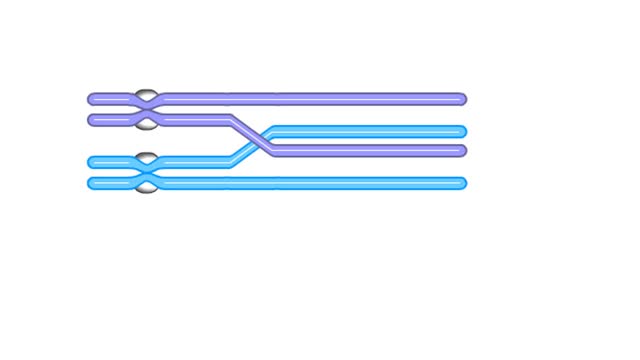
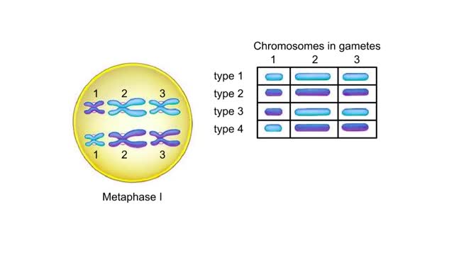
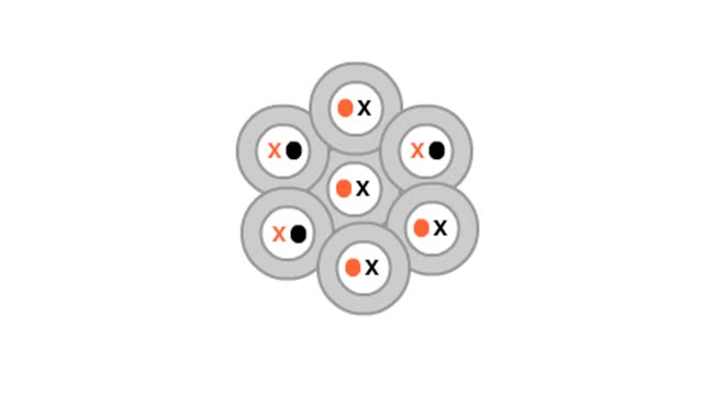
How the chromosome is organized. At metaphase, the chromosomes are duplicated and are at their most condensed. In each chromosome, two identical sister chromatids are held together at a constricted region called the centromere. When a chromosome is condensed, interactions among chromosomal proteins keep loops of DNA tightly packed into a "super coiled array. On closer examination, we can see that the chromosomal proteins and DNA are organized as a cylindrical fiber. If we immerse a chromosome in saltwater. this fiber loosens up to reveal a beads-on-a-string organization. The string is one molecule of DNA. Each bead is a nucleosome that consists of a DNA molecule looped around a core of histones. Nucleosomes can loosen in controlled ways to give enzymes access to specific DNA regions. At mitotic metaphase, the fully-formed spindle is composed of many microtubules that extend from the poles. Some of these, the kinetochore microtubules, are attached to the kinetochores of each chromosome. Kinetochores are located at the centromeres. At anaphase, sister chromatids separate and are pulled to opposite poles of the cell. During this chromosomal movement, the kinetochore microtubules become progressively shorter. The shortening occurs because the kinetothore disassembles the microtubule into tubulin protein subunits as it passes. A motor protein actively "walks" the kinetochore along the microtubule. The tubulin subunits are reused for later microtubule assembly. The following experiment shows that the kinetochore microtubules are shortened by disassembly rather than by contraction or movement toward the pole. Researchers used a microscopic beam of ultraviolet light to "bleach" a section of the microtubules. As the chromosomes moved toward the pole, the bleached segment did not move, indicating that the microtubules themselves do not move. More recent experiments suggest that kinetochore microtubules may also be disassembled at the poles, contributing to their shortening. In contrast to the kinetochore microtubules, nonkinetochore microtubules of the spindle (those not attached to chromosomes) do move, causing the spindle to lengthen and pushing the poles apart. The lengthening of the spindle contributes to the movement of chromosomes away from each other. In the zone where microtubules from different poles overlap, they are pushed apart by motor proteins "walking" between adjacent microtubules.
Add To
You must login to add videos to your playlists.
Advertisement
Suggestions
Comments
0 Comments total
Sign In to post comments.
No comments have been posted for this video yet.